L1- Cardiovascular System (HEART)
CARDIOVASCULAR SYSTEM
It is a closed system composed of the heart and blood vessels.
Blood vessels are
- arteries
- arterioles
- capillaries
- veins

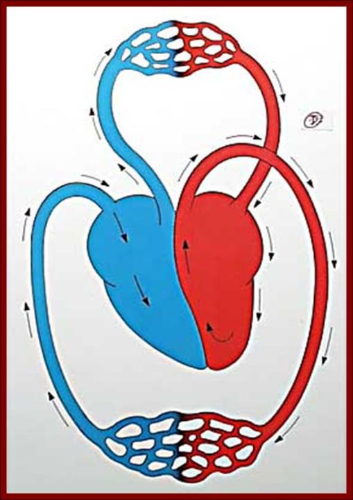
DIVISION OF CIRCULATION
A) SYSTEMIC CIRC.
LV → aorta& branches → arteries → arterioles → capill. → SVC & IVC →RA → RV.
B) PULMONARY CIRC.
RV → Pul art. & branches → rt & lt lungs → capill (gas exchange) → 4 pul veins → LA → LV.
PHYSIOLOGICAL ANATOMY OF THE HEART
It is a hollow muscular organ.
Lies in the left side of the thoracic cavity.
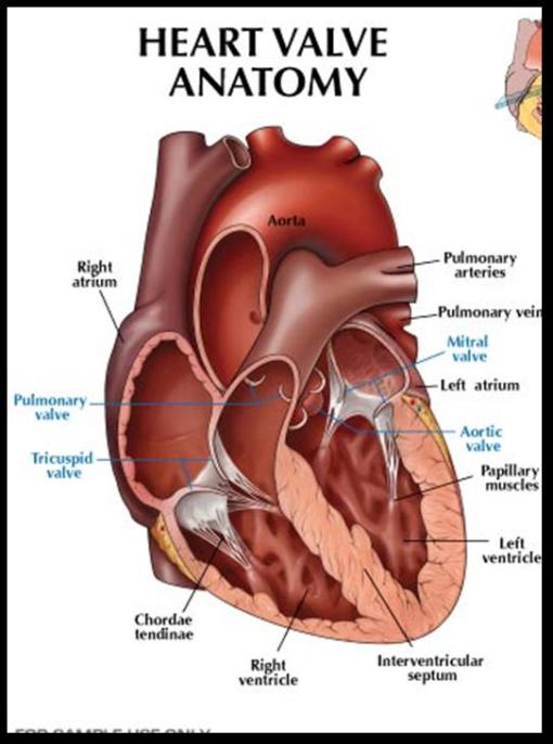
Composed of 4 chambers : 2 atria & 2 ventricles
Enclosed within a pericardial sac which allows optimal distension for the heart.

Cardiac muscle fibers
1.Nodal fibers
- SAN→initiates the c.impulse
- AVN →under conditions
2.Conducting fibers
- AV bundle & its branches
- Purkinjie fibres
3.Contractile ms fibers
a.Atrial ms
b.Vent. Ms
•thicker than atrial ms
•LV ms is times thicker than RV for powerful contraction
Function of the Atria
Blood reservoir on both sides
Pumping of 30% of blood from atrium to vent.
Receptors for CV reflexes
AVN & SAN for conduction of cardiac impulses.
Function of the ventricles
Pumping the blood into the circulation against resistance in big arteries.
Cardiac valves
2 atrio-ventricular valves AVV:
- tricuspid valve between RA & RV
- mitral valve between LA & LV
2 semilunar valves:
- aortic valve between LV & aorta
- pul valve between RV & pul artery

Cardiac Muscle (Myocardium)
Cardiac ms is a striated muscle
Cell membrane separating the cardiac ms cells is called intercalated disc or gap junction
Electrical resistance through them is very small ;so the cardiac impulse travels easily to all C ms.
All cardiac ms act as one unit (functional syncytium)
- The 2 atria → one syncytium
- The 2 vent → one syncytium

Cardiac properties
All are myogenic
- Rhytmicity
- Excitability
- Conductivity
- Contractility
1.Rhythmcity
Ability of the heart to beat regularly on its own
-SAN: 110 bpm
-AVN: 90 bpm
-AV bundle: 45 bpm
-Purkinji fibers: 35 bpm
-Vent.: 25 bpm
Rhythmicity of the heart is a self excitation process that causes automatic rhythmic contraction.
SAN has the greatest automatic rhythm
It is the pacemaker of the heart
It controls the rate of the whole heart

AP of diff parts of the heart
2.Excitability:
Is the ability of the cardiac ms to respond to stimuli
Excit. Changes during AP
a.Absolute reractory period(ARP)
Excit of cardiac ms is lost.
No excitation occurs in it.
It coincides with the systole
b.Relative refractory period (RRP):
Excit gradually recovers
A stimulus can produce weaker systole
Coincides with the 1st half of diastole
c.Super normal phase (SNP):
Excit rises above normal
A weaker stimulus is needed .
Occupies the 2nd half of diastole.
3.Conductivity:
It is the ability of the c.ms to conduct a cardiac impulse through the heart.
1-Atrial transmission:
From SAN -à AVN through atrial ms
2-AV nodal transmission:
Impulse is delayed at the AVN
3-Purkinji system transmission:
From AVN -à AVB & Purkinji fibers.
Very rapid.
4-Vent ms transmission:
To the ventricular ms fibers.

4.Contractility:
It is the ability of the cardiac ms to contract
Contractility depends on:
A-All or none rule:
cardiac ms contracts maximally or does not contract at all ,provided that all the conditions remain constant.

B-Staircase phenomenon:
Cardiac ms shows gradual increase in contraction following a rapid repeated stimulation

C-Starling law:
Wthin limit, the greater the initial length of c.ms the greater the force of cont.

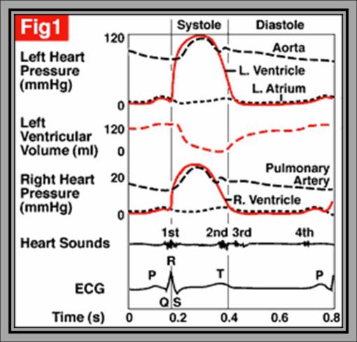
Cardiac cycle
It consists of contraction (systole) followed by relaxation (diastole) of the heart .
1.Atrial systole: 0.1 sec
-atrial p ↑
-is pumping blood from A to V through opened AVV and P retuns to normal

2.Ventricular systole: 0.3 sec
-AVV closed & vent. contract isometrically.
-Vent P ↑
-Ao valve is opened & blood is pumped from LV → aorta at high velocity
3.Ventricular diastole: 0.5 sec
-semilunar valves are closed
-Vent relax isometrically
-Vent P is much decreased to zero.
-AVV are opened
-fall of blood from A to V by its weight

Remarks on CC:
Duration of CC =0.8 sec
- At syst = 0.1 sec
- At diast = 0.7 sec
- Vent syst = 0.3 sec
- Vent diast = 0.5 sec
Vent filling
- 30% in AT systole – active
- 70 % in Vent diastole – passive
Pressures in CC
- Syst BP in LV = 130mmHg
- Dias BP in LV = zero
- Syst BP in RV = 35
- Diast BP in RV = zero
- Syst BP in aorta = 120
- Diast BP in aorta = 80
- Syst BP in Pul art = 30
- Diast BP in Pul art = 10


