L3-Structure of The Cell

THE CELL
- The structural and functional unit of life.
- The smallest unit that display the characteristics of life, i.e. reproduction, metabolism, response to stimuli
- formed of a complex structure called protoplasm.
Cell Characteristics
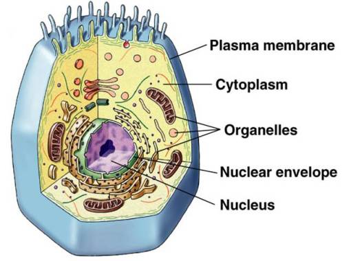
•Plasma membrane
- selectively permeable boundary between cell and environment
•Genetic material ( Nucleus )
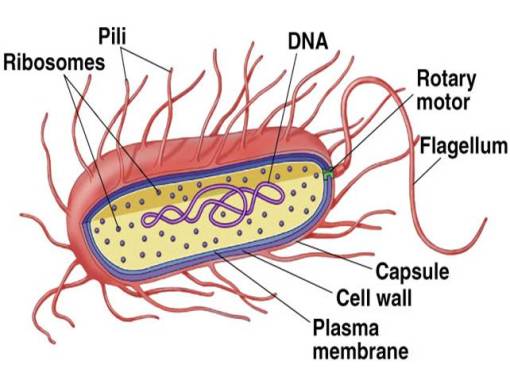
- single molecule of DNA in prokaryotes
- double helix of DNA in eukaryotes
•Cytoplasm
- everything between plasma membrane and nuclear compartment
- fills cell interior
Generalized Eukaryotic Cell
 Prokaryotic Cells

THE CELL MEMBRANE
(Plasmalemma or Plasma Membrane)
•outer limiting membrane
•surrounds the cell
•regulates the passage of materials into or out of the cell.
LM:
•It is very thin to be resolved.
EM:
•three layers (trilamellar)
•outer and inner dark (electron dense) layers
•A middle light (electron lucent) layer

Structure
- phospholipid bilayer
- proteins embedded in, and attached to, the inner (intracellular) and outer (extracellular) surfaces
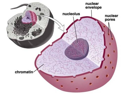
Nucleus
•Repository for genetic material
•Usually single / some cells several / RBC none

1.Nuclear Envelope (membrane)
a.Phospholipid bilayer with nuclear pores – protein gatekeepers
b. Controls what enters/leaves the nucleus
— things only go in or out by passing through
• protein channels, which are selective • Usually proteins going in and RNA going out
c. Encloses all the chromosomes
2.Chromatin
= all the chromosomes, which are long strands of the molecule DNA
— DNA regulates all cell activities, yet never leaves the nucleus; how is this possible?
–produces RNA, short messenger molecules
that exit through nuclear pores
–RNA carries instructions out into the cytoplasm
3.Nucleolus
a. compartment in the nucleus where ribosomes
are assembled
b. ribosomes are then moved out into cytoplasm
through nuclear pores
c. ribosomes and RNA work together outside
the nucleus, to build all the proteins in the cell
THE CYTOPLASM
- Cell organelles
- Cell inclusions
- Cell matrix
1) THE CELL ORGANELLES
•living components in the cytoplasm
•essential for life of the cell
•perform specific functions inside the cell.
Types of Cell Organelles
1- membranous cell organelles
2- non membranous cell organ
1-Membranous Cell Organelles
•surrounded by a unit membrane
•important for the metabolic activity in the cell.
1.mitochondria
2.Golgi apparatus
3.Lysosomes
4.endoplasmic reticulum (smooth and rough)
5.peroxisomes
6.coated vesicles
2-Non-membranous cell organelles
•not surrounded by unit membrane
•important as a cyto-skeleton of the cell
1.Microfilament
2.Microtubules
3.Centeriole
4.cilia and flagella
1) Mitochondria
-Mitochondrion = “thread granule”
-(power house of the cell)
a. energy is taken from sugar, stored ATP
b. requires oxygen to make (aerobic metabolism)
c. contained within double membrane
-Localization of the Mitochondria in the Cell
At sites of high-energy requirement in the cell:
- apical part in ciliated cells
- basal part in of protein synthesizing cells
- between myofibrils in skeletal muscle.
 2) Ribosomes
•non-membranous cell organelles
•Ribosomes = site of protein synthesis
–assembled in the nucleolus
–exported into the cytoplasm
•Ribosomes are RNA-protein complexes composed of two subunits that join and attach to messenger RNA
 Types of ribosomes:
a.Free
- unbound in the fluid cytoplasm
- produce proteins for use in the cell
b.Bound
- attached to the (ER)
- produce proteins for export, or for the plasma membrane
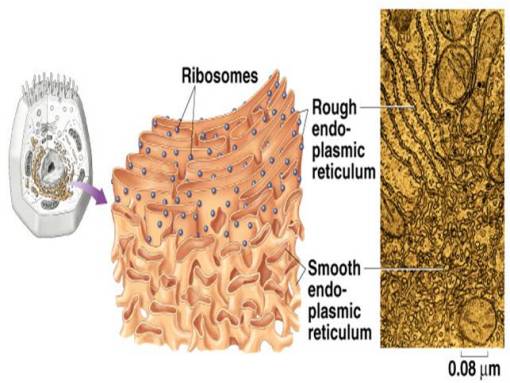
3) Endoplasmic Reticulum (ER)
•= “within the cytoplasm network”, a system of tubes and sacs formed by membranes (an enclosed space)
•Largest internal membrane
•Composed of Lipid bilayer
•Serves as system of channels from the nucleus
•Functions in storage and secretion
•Types of ER
- Rough ER – studded with ribosomes
–modifies proteins produced by ribosomes
- Smooth ER – few ribosomes
–doesn’t modify proteins
–functions in
lipid synthesis
drug detoxification
carbohydrate metabolism
 4) Golgi apparatus
-collection of Golgi bodies
•collect, package, and distribute molecules synthesized at one location in the cell and utilized at another location
•Front – cis , Back – trans
•Cisternae – stacked membrane folds


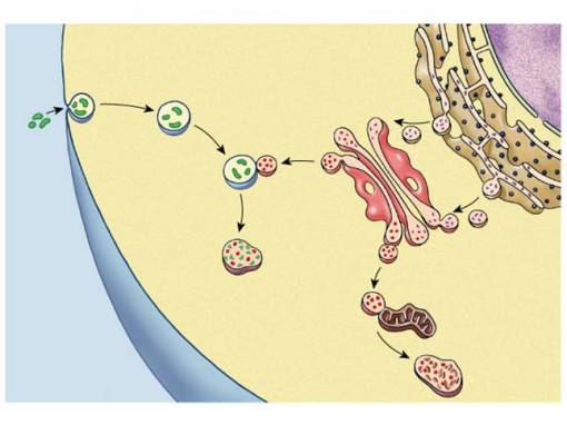
 5) Vesicles
- Lysosomes
•membrane-bound vesicles
•containing digestive enzymes – from Golgi
- Microbodies
•enzyme-bearing, membrane-enclosed vesicles.
•Peroxisomes – contain enzymes that catalyze the removal of electrons and associated hydrogen atoms
 6) Cytoskeleton
•Network of protein fibers
•supporting cell shape
•anchoring organelles
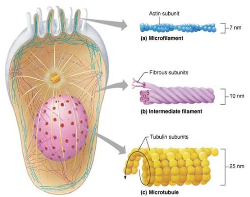
•Cytoskeleton Components
- Microfilament
- Intermediate Filament
- Microtubule
 1-Thin filaments
•Actin micro-filaments (6-7 nm)
- contractile filaments
- inter-act with myosin
•Site
- micro-villi
- cleavage furrow
- muscles
2-Thick filaments
•myosin (12-16 nm).
•thicker than thin filaments
Site:
•In muscle in association with actin filaments forming the myofibrils.
3- Intermediate Filaments
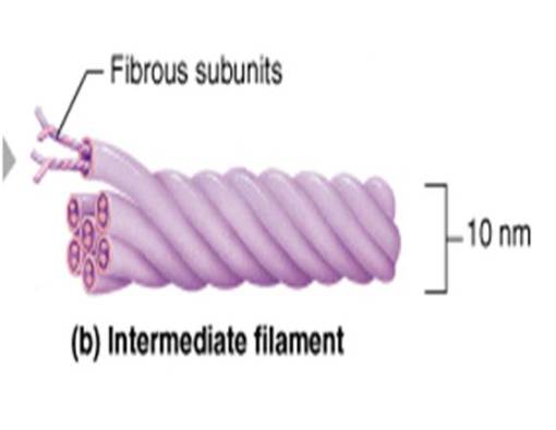
•Tough, insoluble protein fibers with high tensile strength.
•The most stable of the cytoskeletal elements
•Act like internal ‘guy wires’ to resist pulling forces on the cell Important for diagnosis of the tumor
•They are not capable of producing contraction.
1.Desmin filaments
2.Tonofilament
3.Vimentin filaments
4.Neuro-filaments
5.Glial filaments
 4- MICROTUBULES
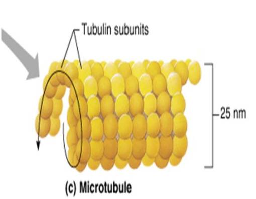
•They are pipe-like structure
•unfixed length
•uniform diameter.
• formed of a protein molecule; tubulin
Distribution:
All over the cytoplasm.
Functions:
1- component of the cytoskeleton supporting, maintaining and stabilizing the shape of the cell.
2- maintain the asymmetrical shape of the cell.
3- the main structural component of cilia, flagella and centeriole.
4- guiding tracks for transporting material and organelles.
7) CENTROSOME AND CENTERIOLES
•The centrosome
- It is a specialized zone of the cytoplasm
- contains two centerioles oriented at right angle to each other.
 •The Centeriole
- It is a non-membranous cell organelle
- important for cell division.
Site:
• in juxtanuclear position in association with Golgi apparatus
2. CELL INCLUSIONS
•non living materials in the cytoplasm.
•products of metabolism or substances that are taken inside the cell from its surrounding.
•types:
1- Stored food.
2- Pigments.
3- Crystals.
1-Stored food
a)Carbohydrates (glycogen granules)
1- alpha glycogen particles
2- beta glycogen particles
b) Fat:
•small droplets large globules
2-Pigment
•coloured substances • seen in the cell without staining.
a) Exogenous pigments:
- Lipochromes pigments
- Dust
- Minerals
- Tattoo marks
b) Endogenous pigments:
- Haemoglobin and its Derivatives
- Melanin
- Lipofuscin pigment


